Haglund deformity: Causes, diagnosis & treatment by foot specialist
- What causes Haglund's deformity?
- Symptoms of Haglund's deformity
- Diagnosis: How does the physician examine Haglund's deformity?
- Remission of Haglund's deformity with conservative treatment
- Surgery for Haglund's deformity
- Important considerations after surgery
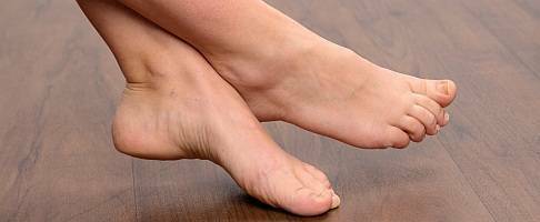
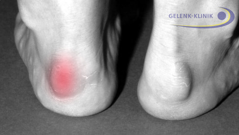 Haglund's syndrome is a deformity of the heel bone (calcaneus) which is also noticeable from the outside. © Gelenk-Klinik
With Haglund's deformity (Haglund's syndrome), part of the heel bone is severely enlarged outward, forming a so-called growth. The Achilles tendon is often irritated or calcified.
The heel in patients with Haglund's deformity often becomes inflamed due to pressure inside the shoe and where the tendons attach to the bone and bursae can be irritated and swollen.
Especially patients with hollow feet (pes cavus) are afflicted with the painful Haglund's syndrome. Stabbing pain in the heel develops suddenly. The foot specialists at Gelenk-Klinik have a number of possible conservative and surgical treatment options. The stage of the Haglund's syndrome determines a suitable treatment.
What causes Haglund's deformity?
Triggers and risk factors of Haglund's syndrome
- intensive running on hard ground,
- tight footwear,
- damage to the talotarsal joint,
- deformity of the calcaneus,
- torsion of the Achilles tendon,
- hollow foot,
- genetic predisposition,
- middle age,
- overweight.
One common cause for pain in the upper heel due to Haglund's syndrome is hollow foot. In this case, the longitudinal arch of the foot is more pronounced than normal so that only the forefoot and heel touch the ground. This results in an altered position of the heel bone (calcaneus) which bulges to the back.
A hereditary protrusion of the heel bone can also irritate soft tissue such as the bursae or the Achilles tendon in the heel. Other causes for Haglund's syndrome are strain from intensive training and tight shoes constricting the foot at the front. Even poorly fitted shoes where the foot can slide can greatly increase stress on the heel when walking. Particularly important for runners: Running on even, hard ground causes particular strain on the heel. Running uphill causes particular strain on the heels. Socks with soft heel cushions can alleviate the strain. Shoes should fit correctly at the heel. Then a severely bulging heel can then remain symptom-free.
Overweight patients who stand a lot at work particularly put a lot of strain on the heel. Overweight people, therefore, increase the chance of developing painful Haglund's syndrome. Conservative treatments have shown little success in overweight patients. The high basic load makes the condition tough and prolongs the treatment for Haglund's syndrome.
Symptoms of Haglund's deformity
The bony growth on the heel bone causes pressure points in the heel area from shoes. Irritations develop both on the Achilles tendon as well as the bursa between the Achilles tendon and heel bone (bursa subachillea).
The painful friction in the shoe causes severe pain in the foot. These can initially arise as pain on initial movement and later also be chronic when walking. Haglund's syndrome can affect one or both feet at the same time.
Pain in the heel may decrease with movement. Overambitious athletes use this effect by pushing "through" the pain during training. This is quite harmful, as the stress and irritation in the soft tissue continue to increase.
Diagnosis: How does the physician examine Haglund's deformity?
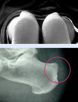 In patients with Haglund's syndrome the heel bone bulges noticeably. The X-ray shows the growth on the heel. Where the Achilles tendon attaches to the bone is already calcified due to irritation. © Gelenk-Klinik
The foot specialist will first assess the appearance and mobility of the foot during a medical examination. The bulge in Haglund's syndrome is typically visible to the naked eye. Patients often also report severe heel pain and problems walking, which provides the physician with additional clues.
An X-ray verifies the visual diagnosis. The lateral X-ray of the foot shows a marked bony protrusion (growth or exostosis) in the heel area. Possible calcifications in the Achilles tendon due to the irritation are also visible in the X-ray. Ultrasound and MRI magnetic resonance tomography help further assess the condition of the soft tissue such as tendons and bursae. A possible inflammation of the bursa (bursitis) is immediately visible.
To determine the stage of the condition, the physician will further determine the so-called Fowler-Philip angle. This angle is significantly greater in Haglund's syndrome. It is read using the lateral X-ray of the foot bearing weight. An angle over 75 degrees indicates Haglund's syndrome.
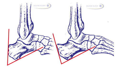 The Fowler-Philip angle is determined using the lateral X-ray of the foot. Left: a small Fowler-Philip angle indicates a healthy heel bone position. Right: The Fowler-Philip angle is significantly greater in Haglund's syndrome. © Gelenk-Klinik
Remission of Haglund's deformity with conservative treatment
Conservative treatment:
- taking a break from training, reducing strain
- insoles to raise the heel by about 1 cm
- heel cushion
- thermotherapy (cold, heat)
- ultrasound treatment
- anti-inflammatory gels or creams
- shockwave therapy
- losing weight
- physiotherapy
- stretches
Treatment is based on the stage of Haglund's syndrome. Relief will alleviate the pain in the heel temporarily. Lasting improvement can only be achieved by correcting the structure. This can for example be orthopaedic insoles which will slightly correct the position of the heel bone. Insoles will further provide relief for the muscles pulling on the Achilles tendon. Another important measure to relieve the heel is for an overweight person to lose weight.
Applying cold or heat or ultrasound therapy also provide good results. Shockwave therapy has been used very successfully at Gelenk-Klinik. If it is determined the Achilles tendon is already damaged, the orthopaedist will consider surgery.
Surgery for Haglund's deformity
There are a number of surgical options to treat Haglund's syndrome and possible damage to the Achilles tendon, ligaments or deformity of the heel bone.
Minimally-invasive surgery for Haglund's deformity
If it is determined the Achilles tendon is only mildly altered due to inflammation, the foot specialist can perform an endoscopy for Haglund's deformity through a small incision with X-ray guidance. Similar to open surgery, with this procedure the bone is ablated from the heel bone, any inflamed bursae and tissue is removed and the Achilles tendon is treated endoscopically.
Advantages of minimally-invasive surgery for Haglund's deformity
The orthopaedic specialists at Gelenk-Klinik check for each patient if endoscopic minimally-invasive surgery can be used instead of open surgery. The extensive experience of the surgeons has shown the following advantages of minimally-invasive Haglund's surgery:
- small scars which do not extend across the entire length of the heel causing irritation, pressure and heel pain when wearing shoes
- minimal damage to tendons and bursae
- quick healing and regeneration, sutures removed after only 7 days
After-care for minimally-invasive Haglund's deformity surgery
A successful outcome of surgery requires immobilizing the foot in a brace. The Achilles tendon must be fully healed before the respective foot can make a full foot rollover. This is about 6 weeks. Extensive rest and partial weight-bearing promote healing.
Open surgery for Haglund's deformity
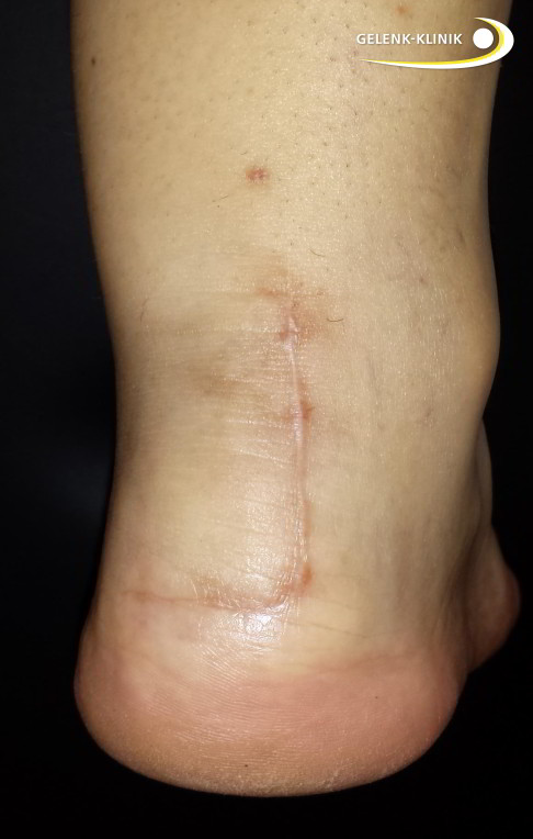 Patient after open surgery for Haglund's deformity. The skin incision is healed. © Dr. Thomas Schneider, MD
If the enthesis is damaged (tendinopathy), open ablation of the bone growth with opening the Achilles tendon and treating the damage to the tendon is the standard procedure. Removing the bone relieves the Achilles tendon. An inflamed bursa can also be removed during this procedure. The open access provides a direct view of the Achilles tendon and the surgeon can also assess its condition. The Achilles tendon is treated if necessary.
After-care for open surgery of Haglund's deformity
During the first few days after the procedure, the patient should rest and immobilize the operated foot. Forearm crutches are helpful in relieving strain. After that, the foot can bear full weight.
Physiotherapy during the first months following surgery help build muscle function and contribute to maintaining physiological mobility. This improves the outcome of surgery and reduces the rate of complications, thus the risk of another procedure (revision).
Stabilizing the Achilles tendon
If the enthesis of the Achilles tendon is severely damaged or degenerated, the Achilles tendon will also need to be treated. The damaged areas of the Achilles tendon are removed and the healthy sections are reinforced with sutures. Even spurs on the Achilles tendon enthesis require sutures: these are very difficult to treat through surgery. It requires detaching part of the Achilles tendon enthesis and reattaching it after the spur has been removed. Suture anchors are used for this and the tendon is sutured tight above this.
Zadek surgery: correction of the heel bone
Removing the bone growth is only one treatment option. Depending on the shape of the heel, an osteotomy which is gentle on the sensitive enthesis may be a good option. This procedure is called Zadek surgery. At Gelenk-Klinik the foot specialists consider a heel osteotomy according to Zadek for the following indications:
- The patient's heel is severely widened and the Achilles tendon enthesis is chronically irritated.
- The X-ray in the standing patient shows a steep heel bone position.
- Due to the deformity in the heel bone, there are comorbidities in the tendons and ligaments.
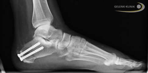 X-ray following Zadek surgery: The corrected position of the heel bone is fixed with screws until fully healed. © Dr. Thomas Schneider, MD
Zadek surgery process
During the osteotomy, a bone wedge is removed from the heel bone and the deformity is permanently corrected. The tendon is therefore permanently relieved. The Zadek surgery offers considerable advantages for some patients. In principle, the Zadek surgery is an open surgery, however, since 2014 the specialists at Gelenk-Klinik have been performing more of these procedures endoscopically.
Advantages of Zadek surgery
The advantages of minimally-invasive correction of the heel bone are obvious: hardly any scars on the heel to cause problems in shoes, less soft tissue injury and faster healing.
Parallel to correcting the bone, the surgeon can remove inflamed bursae, repair the Achilles tendon or block painful nerves. The Zadek surgery is tailored to each patient and requires the treating orthopaedist to have a lot of experience. Gelenk-Klinik guarantees this with its certification as a Foot and Ankle Surgery Centre (ZFSmax).
When can I return to sports and drive again?
Based on experience, the calcaneus heals very well, and the Achilles tendon attachment may still be swollen and sensitive to pressure for some time. Complications such as infections, damage to vessels, nerves, ligaments, tendon irritation and having the surgery repeated are possible, but very rare compared to other surgical methods.
Driving is not permitted until full weight bearing is reached. Most patients can start low-impact sports such as biking and swimming after 6 weeks. Activities that strain the foot such as walking and running can only be resumed after 12 weeks. Always consult the physician before any planned activities.
Important considerations after surgery
Immediately after surgery you should elevate and ice your foot to minimize pain and swelling. The sutures will be removed after about 10 days. At that point, you may shower again.
You will also be using a special shoe. For longer distances, you should also use forearm crutches to relieve your foot. It’s vital to use thrombosis prophylaxis until your foot returns to full weight-bearing.
Fact sheet minimally-invasive Haglund's surgery
- Inpatient treatment: 2 days
- Mobilization: Full weight bearing in VACOped orthosis for 2 weeks
- Earliest flight home: 5 days after surgery
- Recommended flight home: 7 days after surgery
- Time before showering: 12 days
- Recommended time off work: 2-3 weeks
- Time before removal of sutures: 12 days
- Time before driving: 6 weeks
- Sport activities: earliest after 3 months
- Outpatient rehabilitation treatment recommended: 2 weeks lymphatic drainage only, after that reactivation and training allowed
Fact Sheet Haglund's surgery – Zadek technique
- Inpatient treatment: 4 days
- Mobilization: Partial weight bearing of 20 kg in VACOped orthosis for 6 weeks
- Earliest flight home: 10 days after surgery
- Recommended flight home: 14 days after surgery
- Time before showering: 12 days
- Recommended time off work: 6-8 weeks
- Time before removal of sutures: 12 days
- Time before driving: 12 weeks
- Sport activities: earliest after 3 months
- Outpatient rehabilitation treatment recommended: lymphatic drainage and passive mobility training for 6 weeks, training can be increased once the orthosis has been removed
