Diagnosis of Knee Cap (patella) dislocation
Diagnostics of patella dislocation
- X-Ray
- Clinical investigation
- detailed case history
- Electromyography (EMG)
- Diagnostic Arthroscopy
- MRI
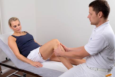
The basic requirements for the successful treatment of kneecap dislocation are a complete case history and a thorough physical examination.
X-rays of the knee joint may provide indications of possible predisposition or existing injuries to the bones. A spot film of the patella, taken while the knee is bent, helps track the extent of patella misalignment. With this information, it is possible to make an estimate of the severity of the chondropathy (cartilage damage) within the knee.
If the clinical findings are ambiguous, an examination under anaesthesia may be recommended or an arthroscopic diagnosis may be necessary.
 Patella dislocation with the free articular body is visible in the left joint. This kind of patella (knee cap) diagnostics requires the knee specialist to have experience in x-ray diagnostics. © joint-surgeon.com
During an arthroscopy it is also possible to remove detached bone or cartilage fragments or to smooth damaged cartilage.
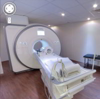
Magnetic Resonance Imaging (MRI) examinations (just like the electromyography (EMG) test checking the health of the leg muscles and nerves), help to establish a more precise and complete diagnosis of the condition of the knee cartilage and soft tissue.


 Prof. Dr. med. Sven Ostermeier, Consultant for Orthopaedic Knee Surgery

